Physician Webinar: The Role of Liver Imaging in HHT (Recorded)
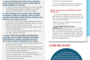
Liver involvement occurs in approximately 75% of people with HHT. While many people are asymptomatic, the complications associated with liver involvement have historically been under diagnosed. These complications include arterioportal shunting leading to portal hypertension and arteriosystemic shunting leading to high output cardiac failure (HOCF). HOCF occurs much more frequently than has previously been considered. The increased recognition of liver vascular malformation and its consequences is noted in the second international HHT guidelines which recommend liver screening for patients with known or suspected HHT. Imaging modalities currently utilized in the screening and diagnosis of these disorders include ultrasound, computed tomography and magnetic resonance imaging (MRI).
Most patients are diagnosed with HOCF after they have already developed symptoms. Current diagnostic considerations look at the secondary effects of shunting on the heart. However, newer imaging techniques allow for the quantification of blood flow through the liver. Such studies could potentially be used one day to predict risks for heart failure before it occurs and a patient becomes symptomatic. They may also be useful to monitor effects of treatment. MRI imaging to measure flow through the liver utilizes protocols and software that are widely available to most imaging centers.
Learn how newer developments in liver imaging may advance the care of liver treatment in people with HHT.
Objectives:
- Know the types of vascular shunts that occur in the liver with HHT.
- Explain how the type of vascular shunting connects to the patient’s symptoms.
- Understand the roles of various diagnostic imaging modalities in following patients with HHT.
Presenters: Mark Ohliger, MD, PhD, Mark Sugi, MD and Kevin Whitehead, MD
Related Resources

HHT International Clinical Guidelines (2020)
My HHT Liver VM Care Checklist