Little Eleanor’s First Center Visit
Wow! What a week it has been! We just got back to Kansas from our trip to the Washington University HHT Center to have our daughter screened for cerebral AVMs.
A reminder for my friends and family who are reading along – an AVM is an arteriovenous malformation that can occur in the organs of patients who have HHT. These blood vessel malformations are susceptible to rupture, causing severe bleeding.
In this post, I will talk about how to initiate a pediatric screening, what happens along the way, what our experience was like experience, and highlight the HHT center for its excellent work.
Preparing
If you are an HHT family considering having children, it’s good to start thinking about pediatric screenings for your child after he or she is born. Make note to ask the delivery team to save blood from the umbilical cord to send for genetic testing and to request meeting with a genetic counselor during pregnancy to discuss HHT, facilitate genetic testing and coordinate meetings with the HHT center of your choice.
Once one person in the family is genetically diagnosed, it makes it easier for everyone else to be tested. Instead of analyzing all the different genes that might cause HHT, the testing will be isolated to one gene, which makes it easier (and cheaper). Eleanor’s grandmother was tested first and her results isolated the endoglin genetic mutation, so Eleanor was only tested for that specific gene.
After your genetic diagnosis is received (or if your child has family history of HHT, nosebleeds, telangiectasias and/or AVMs), pediatric screening at an HHT center is highly recommended. Young children typically do not experience nosebleeds or telangiectasias, but that does not mean they are free of AVMs in major organs.
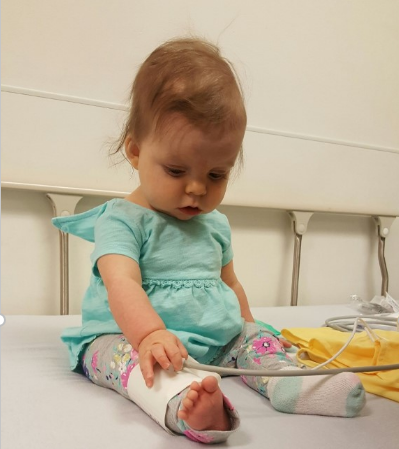
Little Eleanor analyzing the blood pressure cuff.
Making the Appointment
The HHT centers operate with a multidisciplinary team of healthcare providers. They can be comprised of specialists in pediatrics, radiology, rheumatology, cardiopulmonary medicine, genetics and many others. This makes for some special treatment for your little one!
There are 25 HHT centers in the US. We live in central Kansas and there are three centers within a six hour drive. I found these centers on the Cure HHT website and contacted each one to chat! Don’t hesitate to call – they are staffed with wonderful and very knowledgeable people who are ready to help you make the best choice for your family.
Here is what I asked:
- What is your standard of care for pediatric screening?
- What tests do you do?
- Do you require a genetic diagnosis first?
- What if the tests are positive?
- Who are the healthcare providers that work in the center?
- How long willthe visit last?
- Will you communicate results with the primary care doctor?
- What is your plan for follow-up?
The University of Texas – Southwestern HHT Center is where my mother-in-law receives her care, but they primarily see adults. I was more comfortable going to an HHT center that deals with children on a regular basis (because in my experience, children are not just mini-adults – they are a whole different kind of beast!). Denver saw children with HHT, but the appointments would have been stretched out over multiple days.
It worked out logistically for us to visit St. Louis Children’s hospital. They were able to get Eleanor in for testing and her clinic appointment in one day. The staff was so incredibly nice when setting up these appointments. They worked with me to get referrals from her pediatrician, gave me all their direct phone numbers and told me exactly what would happen and when.
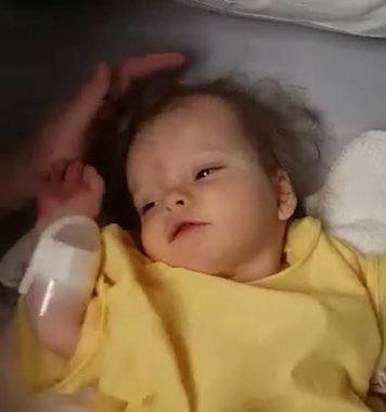
Waking up from anesthesia – no negative side effects, just extra hungry.
Typical Process
A child undergoing screening is sent to radiology/imaging in the hospital early in the morning. Very young children must fast and be anesthetized for the imaging because they cannot get good pictures if your kiddo is being a wiggle-worm. An MRI is done of the brain, taking pictures with and without a contrast agent. The contrast agent is given through an IV and helps improve image quality so doctors can see the blood vessels in the brain better. The scan takes about an hour.
As the child is waking up from anesthesia, they will have a bubble echocardiogram to check for pulmonary AVMs. An echocardiogram is basically an ultrasound of the heart. The bubble part comes in when they inject saline and tiny air bubbles into the IV and watch for the bubbles on the echocardiogram. Normally, the lungs will filter out the bubbles as the blood circulates through them and back to the heart, or the bubbles will appear after several heartbeats because they will follow the normal circulatory path.
If an AVM is present in the lungs, the bubbles will appear in the left side of the heart much sooner (after 3-5 heartbeats). This means the bubbles went into the lungs and back to the heart quickly because they bypassed capillaries, which normally would have slowed them down or filtered them out.
A positive bubble echocardiogram means an AVM is suspected, but to confirm this and the size/number/location of AVMs, a CT scan of the lungs is typically done next. The CT scan is not done initially because it involves exposing the child to radiation.
Getting acquainted with the Washington University HHT Center Team.
Our Experience
I woke Eleanor up in the middle of the night to nurse her, as she couldn’t have any solids, including breastmilk, after 2 a.m. In the morning, she was very hungry and cranky! Honestly, the hardest part of the entire day was not being able to feed her when she was so hungry.
We arrived at the HHT center and were taken back to radiology. The doctors and nurses took her vitals, medical history, and did a great job at keeping us all comfortable and informed. When it was time for the IV, a child life specialist came in with some neat toys to help keep Eleanor distracted. The nurses did an excellent job placing it, and our baby girl was very brave. She then went down to the MRI, and in that room, she was anesthetized and scanned.
When she started waking up after the scan, we were allowed back in recovery to be with her. She was pretty drowsy and silly for a bit, but soon she was ready to eat and get out of there. El has a congenital heart defect that would cause a positive bubble echo, making it impossible to determine if she has pulmonary AVMs by that test, so we decided not to do the echo and to skip the CT scan of her lungs for now. If she has a procedure in the next few months to get her heart fixed, we will use that opportunity to scan her lungs for AVMs.
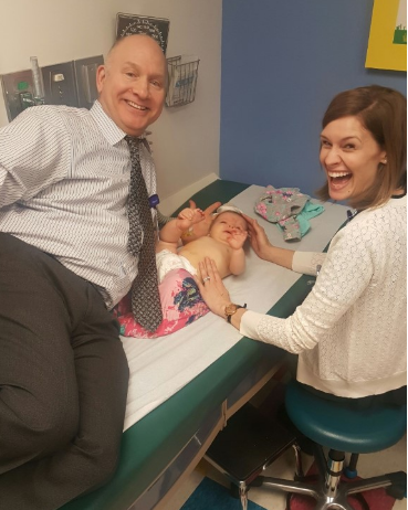
After leaving the MRI, we went to lunch and came back to the center to see Dr. White. Eleanor was weighed, measured and her vital signs were recorded. We then spent time speaking with resident Dr. DeSilva, Dr. White and Nurse Coordinator Lynn Sekarski. They went over our family history, talked to my husband about his symptoms, asked about Eleanor’s history, and gave her a little physical.
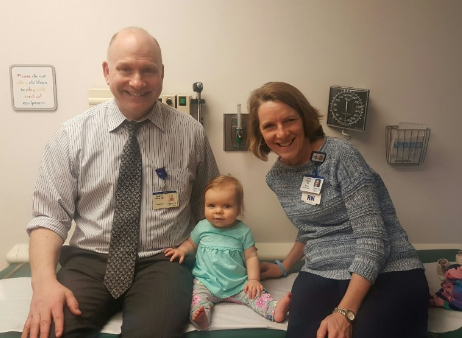
Eleanor with Dr. White and Nurse Coordinator Lynne Sekarski.
Fortunately, Eleanor does not have any AVMs in her brain! We were so excited and relieved to hear this news. Since cerebral AVMs are suspected of only developing during times of rapid growth, she will not need another brain scan until puberty in about 10 years or so. The team at the center was so nice.
My husband said he has never had so much fun at a doctor’s office. I believe, even if we received bad news, the experience would have been the same. Dr. White and Lynn are both very passionate about the work they do within the HHT community. We hope to continue Eleanor’s care with them, even as we move around the country with the military.